Scleroderma
Scleroderma in childhood is rare and has variable clinical presentations. It is broadly divided into localized scleroderma and systemic scleroderma, which differ significantly in severity, complications and management.
Localized scleroderma (morphoea)
Localized scleroderma is the most common form in children and can present at any age. It typically appears as a patch of abnormal, thickened skin (morphoea).
The condition often follows a course of active expansion, fibrosis (skin thickening and hardening), and eventual softening or partial remission.
Despite being confined to the skin and underlying tissues, the functional and cosmetic impact can be significant. Lesions may interfere with growth of a limb, affect subcutaneous tissues of the face or body, and result in asymmetry or deformity.
Systemic scleroderma
Systemic scleroderma is extremely rare in children but is a serious, progressive condition. It is characterised by diffuse fibrosis of the skin and involvement of internal organs including the lungs, gastrointestinal tract, heart and kidneys.
The disease is typically slowly progressive, has a guarded prognosis, and is associated with significant morbidity and mortality.
Management
Management of childhood scleroderma requires specialist multidisciplinary care. Treatment often includes corticosteroids, methotrexate and other immunosuppressive or targeted therapies.
The aim of treatment is to limit disease progression, prevent growth disturbance, and reduce functional impairment and cosmetic complications.
Comparison of localised and systemic scleroderma
|
Feature |
Localized scleroderma (morphoea)
|
Systemic scleroderma
|
|
Frequency |
Most common form in children
|
Extremely rare |
|
Age at onset |
Any age
|
Rare in childhood |
|
Clinical appearance |
Localised patch of abnormal skin
|
Diffuse skin fibrosis with organ involvement |
|
Disease course |
Expanding → fibrosis → softening/remission
|
Slowly progressive, guarded prognosis |
|
Complications |
Growth impairment, facial/limb deformity, cosmetic impact
|
Lung, GI, heart and kidney involvement; significant mortality |
|
Management |
Early recognition and treatment to prevent disability
|
Aggressive immunosuppression and specialist care |
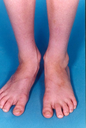
The photograph below shows linear scleroderma with pigmented indurated lesions and the right foot is smaller than the left.
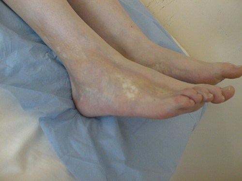
The photograph below shows linear scleroderma with typical 'waxy' pigmented lesions over the foot.