Radiographs (X-Rays)
- Radiographs are usually normal in early inflammatory arthritis (JIA). Although plain radiographs may reveal soft tissue swelling around joints or demonstrate bony erosions, radiographs are an insensitive tool for the diagnosis of acute synovitis or erosive joint damage. Ultrasound scanning or Magnetic Resonance Imaging is preferable.
- Plain radiographs can be used in the initial investigation of children with bone pain or arthritis.
- X-rays are often used to rule out other causes of joint pain, rather than assess for JIA; the presence of periosteal reaction suggests possibility of bony infection or malignancy.
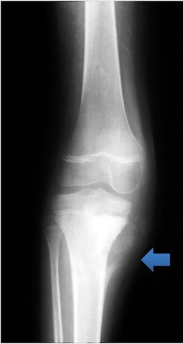
- Radiographs of the knee should include the long bones above and below in the context of persistent knee pain - bone tumours commonly arise in the distal femur or proximal tibia.
- An anterior-posterior view of the hand and wrist can be a useful screening tool in the investigation of metabolic bone disease (e.g., rickets).
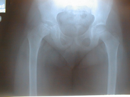
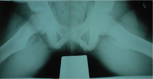
- It is often useful to compare both sides (e.g., hip radiographs) to exclude pathology. Frog-leg views should be requested to detect subtle changes of Slipped Upper [Capital] Femoral Epiphysis.
The Figure below shows an abnormal hip (right) - due to Perthes
The Figure below shows an abnormal hip (right) - compare both sides - due to Slipped Upper [Capital] Femoral Epiphysis
The radiograph below shows periosteal reaction and soft tissue swelling due to malignant bone tumour in the proximal tibia (arrow). This emphasises the need to include images of the long bone above and below the knee when imaging for knee pain.