Juvenile Dermatomyositis
- Juvenile Dermatomyositis (JDM) has a broad range of severity and is a serious disease requiring specialist assessment and management.
- JDM can present at any age, with a variable presentation and course.
- Typical features include:
- Characteristic skin involvement - malar rash, heliotrope rash over the eyelids, photosensitivity, vasculitis, and nail fold capillary changes. Calcinosis (painful or painless lumps or sheets under the skin) are a feature of late diagnosis or poorly controlled disease.
- Chronic skin disease can result in Gottron's papules over the knuckles, elbows or knees. Skin ulceration may also be present.
- Proximal muscle weakness can present acutely or indolently with difficulty on stairs or fatigue on walking.
- Myalgia, arthralgia and arthritis and joint contractures.
- Systemic symptoms such as malaise, fever, weight loss or anorexia
- Bulbar muscle involvement and chest wall muscles weakness may result in risk of aspiration and pneumonia.
- Bulbar muscle involvement and chest wall weakness may be suspected if patients have a weak voice, difficulty swallowing, shortness of breath of difficulty coughing.
- Remember 'dysphagia, dysphonia, dyspnoea'.
- Interstitial lung disease and gut vasculitis can occur with potential for severe morbidity and mortality.
- Unlike adult onset DM, JDM does not tend to be associated with malignancy.
- Routine screening for malignancy in children with JDM is not necessary.
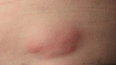
The photograph below shows subcutaneous calcinosis in JDM
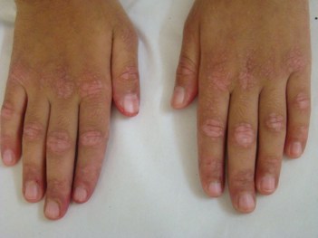
The photograph below shows typical Gottron's papules over the knuckles in JDM
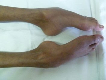
The photograph below shows severe joint contracture and muscle wasting from chronic JDM that has not been treated optimally.
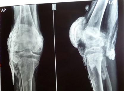
The radiograph below shows severe calcinosis around the knee in chronic JDM
- The diagnosis of JDM relies on clinical assessment and investigations (including elevated serum muscle enzymes, imaging and tests to exclude other differential diagnoses).
- However it is important to consider inherited myopathies which can also cause elevated muscle enzymes; there may not be a family history and any unusual features warrant further investigation which may include muscle biopsy.
- Notably, most children with JDM do not require electromyography or a muscle biopsy except where the presentation is atypical. Electromyogram (now rarely performed) shows myopathy / denervation.
- Magnetic resonance imaging (MRI) of the muscles helps to identify muscle involvement and monitor disease activity; MRI is now regarded as diagnostic.
- Muscle biopsy may show inflammation, fibre necrosis and small vessel vasculitis but is not always required for diagnosis.
- The management of JDM is multidisciplinary.
- Treatment of JDM requires high dose corticosteroids, often given intravenously, frequently accompanied by methotrexate, although other medications such as intravenous immunoglobulin, cyclophosphamide or anti-cytokine (biologics) agents are used in severe or refractory disease.
- Any suspicion of bulbar of chest wall muscle involvement (weakness of speech or cough, difficulty in eating or swallowing or shortness of breath) warrant urgent specialist intervention.
- Speech and language therapist assessment is important to assess the risk of aspiration.
- Physiotherapy and occupational therapy are vital to optimise outcomes and especially in the rehabilitation phase.
- Specialist nurse input is important to support patients and families.
- Progress is monitored with clinical assessment, muscle enzymes and tools to assess muscle power and function.
- Useful resources to demonstrate skin lesions:
- Welcome to Skin Deep - Skin Deep (dftbskindeep.com).
- https://www.nottingham.ac.uk/research/groups/cebd/resources/skin-of-colour/index.aspx.
- Further information about myositis is available.
- More information about the clinical assessment of muscle disease is available.
- More Information about the approach to investigation of muscle disease is available.