Vasculitis
Vasculitis is the term to describe heterogeneous disorders with presence of inflammation of blood vessel walls. Patients with vasculitis can present with various clinical findings based on the size of affected vessels. Vasculitis is suggested by a spectrum of clinical and laboratory features.
When to suspect vasculitis
- Constitutional symptoms (e.g., fever, weight loss, persistent fatigue).
- Multiple organ involvement.
- Pathognomonic signs of vasculitis (e.g., Mononeuritis multiplex, brain infarction, hypertension, pulmonary hemorrhage, stroke in the young, palpable purpura, livedo reticularis).
Step by step approach to suspected primary vasculitis.
Step 1: Exclude vasculitis mimics (e.g., infective endocarditis, anti-phospholipid syndrome, thrombotic thrombocytopenic purpura, paraneoplastic syndrome).
Step 2: Exclude secondary vasculitis.
- Infections: hepatitis, HIV, CMV, mycobacteria, mycoplasma, COVID-19
- Autoimmune: SLE, inflammatory bowel disease, juvenile dermatomyositis
- Malignancies: lymphoma, lymphoproliferative syndrome
- Medication: serum sickness, amphetamines, cocaine
Step 3: Defining the primary vasculitis syndrome.
Step 4: Classify type of vasculitis based on clinical phenotypes and size of affected vessels (Small/Medium/Large) as described in the Table below:
|
Large blood vessels |
Medium sized blood vessels |
Small blood vessels |
|
Claudication |
Subcutaneous nodules |
Palpable purpura |
|
Pulse absence/unequal |
Deep ulcers |
Infiltrated erythema |
|
Bruits |
Livedo reticularis |
Urticarial vasculitis |
|
Asymmetric blood pressure |
Digital gangrene |
Superficial ulcers |
|
Hypertension |
Mononeuritis multiplex |
Glomerulonephritis |
|
|
Erythema nodules |
Pulmonary haemorrhage |
|
|
Hypertension |
Gastric colic |
|
Classification of primary childhood vasculitis |
|
|
Large vessel vasculitis |
Takayasu arteritis |
|
Medium-sized vessel vasculitis |
Polyarteritis nodosa |
|
|
Kawasaki disease* |
|
Small-sized vessel vasculitis |
|
|
ANCA-associated vasculitis |
|
|
- Granulomatous |
Granulomatosis with polyangiitis (GPA); former name: Wegener’s granulomatosis Eosinophilic granulomatosis with polyangiitis (EGPA); former name: Churg-Strauss syndrome |
|
- Non-granulomatous |
Microscopic polyangiitis (MPA) |
|
Other small-sized vessel vasculitis |
IgA vasculitis (Henoch Schönlein purpura)* |
|
Variable-sized vessel vasculitis |
Behcet’s disease |
*Most common vasculitis in children
Abnormal laboratory features may include:
- Increased acute-phase reactants (ESR, CRP).
- Anaemia, Leukocytosis, Eosinophilia (EGPA).
- Antineutrophil cytoplasmic antibodies (ANCA).
- Most (75-95%) patients with GPA are c-ANCA or anti-proteinase 3 Antibodies positive
- Many (40-80%) patients with EGPA, MPA are p-ANCA or anti-myeloperoxidase antibodies positive
- Elevated factor VIII - related antigen (von Willebrand factor).
- Haematuria and active urine sediment.
- Histopathology can be performed in medium or small-sized vessel vasculitis.
- Imaging study (e.g., MRAngiogram, CTAngiogram) may be useful to diagnose medium or large-sized vessel vasculitis. US or CT abdomen may be performed in complicated cases of HSP.
Useful resources to demonstrate skin lesions in vasculitis:
Welcome to Skin Deep - Skin Deep (dftbskindeep.com)
https://www.nottingham.ac.uk/research/groups/cebd/resources/skin-of-colour/index.aspx
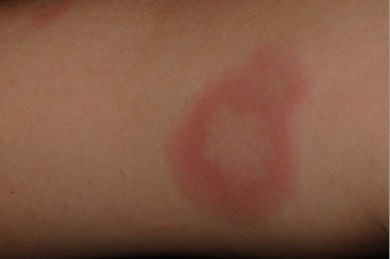
The photograph below shows a 'target lesion' of skin vasculitis
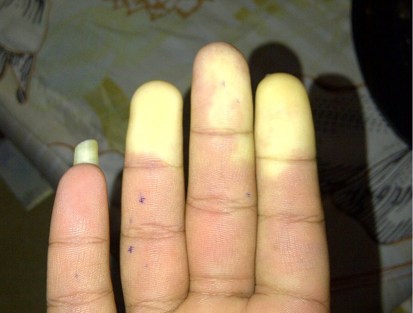
The photograph below shows ischaemic fingers in vasculitis with antiphospholipid syndrome