Malignancy
Childhood malignancies frequently present with musculoskeletal symptoms and signs.
NICE Guidance on when to suspect cancer in children is helpful and includes consideration of musculoskeletal features.
Malignancy should be excluded in all children presenting with arthritis, and should always be excluded before a diagnosis of Juvenile Idiopathic Arthritis (JIA) is made and certainly before starting systemic corticosteroids.
Red flags: fever, systemic features (weight loss, fever, pallor, petechiae, lymphadenopathy, lethargy, malaise, hepatosplenomegaly), bone pain, back pain, nocturnal pain, pain out of keeping with physical findings.
Leukaemia
Leukaemia is the most common form of childhood cancer. The most common form of leukaemia in childhood is Acute Lymphoblastic Leukaemia (ALL), which typically presents around the age of 2-3 years. Arthritis is detected in around 25% at presentation. Distinguishing between leukaemia and JIA can be challenging; there may be overlap of systemic features, such as grumbling fever or hepatosplenomegaly seen in systemic-onset JIA and connective tissue disorders. Investigations include blood tests and imaging; children with ALL may have a normal/low white cell count and normal blood film; diagnostic blast cells are often absent at disease onset. Suspicion is increased with the following; - depression of all three cell lines or at least two series, a differential count with dominant lymphocytes, a raised ESR with elevated LDH and uric acid. Bone marrow aspirate may be needed to confirm or exclude leukaemia.
Lymphoma
Lymphomas less commonly present with musculoskeletal manifestations, but bone pain may be present. There are many features in common with systemic-onset JIA and lymphoma (arthralgia, fever, malaise, lymphadenopathy, splenomegaly), so care must be taken to distinguish between the two diagnoses. Investigations include blood tests and imaging. Lymph node biopsy is required to confirm the diagnosis.
Neuroblastoma
Neuroblastomas are the commonest extracranial tumours in children, most presenting under 5 years of age. Presenting features are usually due to mass effects or metastases. Bony metastases are common and children may report bone pain. Back pain is very unusual in young children, and if reported should be taken seriously.
Bone Tumours
Bone tumours in children may be benign or malignant. Malignant tumours are rare, but serious and potentially fatal, so it is essential to identify them early. Common sites are distal femur and proximal tibia so care must be taken in the case of knee pain that is unusual, not responding to treatment or there are red flags in the presentation. Radiographs of the knee should include the long bone above (femur) and below (tibia / fibula). Malignant tumours include Osteosarcomas (55%) Ewing’s sarcoma (35%) and chondrosarcomas (<5%). Benign tumours incorporate osseous tumours, such as osteoid osteomas and osteoblastomas, fibrous tumours, such as non-ossifying fibromas, and cartilaginous tumours, such as osteochondroma. Typically osteoid osteomas present with night pain and common sites are the spine and long bones. The pain may respond to NSAIDS. Benign bone tumours may be an incidental finding on radiograph - treatment may not be needed; depending on whether the lesion is causing any symptoms.
Any suspicious symptoms or lesions should be referred urgently - orthopaedics, rheumatology or general paediatrics and depending on local referral pathways. Referral should not be delayed by waiting for investigation results. Radiographs will detect most bone tumours; further imaging may be needed (e.g. the site, extent of lesion(s), biopsy guidance, assess for metastases).
Presenting features are common to different types of tumour, and can include:
- Bone pain / night pain
- Swelling of the soft tissue or bone
- Systemic features eg lethargy, weight loss
- Radiographs - sometimes, bone tumours are detected coincidentally, such as following minor injury but may also present with a fracture through the lesion (pathological fracture).
The bone scan below shows 'hot spots' in the pelvis and the right femur due to metastatic bone cancer - bone scans are less frequently used if MRI is available as the latter is without radiation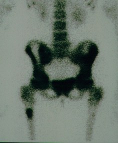
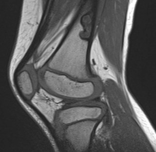
The MRI below shows a benign bone lesion in the distal femur
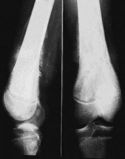
The radiograph below shows a malignant bone tumour in the distal femur (note the soft tissue swelling and irregular cortex)
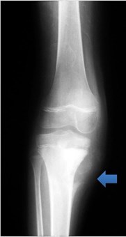
The radiograph below shows the cortex changes and soft tissue swelling (arrow) of osteosarcoma in the proximal tibia